LSM 710 - Confocal Scanning Microscope
Location and contact information
Location: CCNY, Marshak 525
Facility Manager: Jorge Morales
Phone: 212 650 8591
Hours: Open 24/7 to qualified users.
Scheduling calendar: click here
Rules governing the usage of the facility
Fees
External federal users - $85/h
CCNY users - $20/h
Long-running experiments qualify for a 50% discount. See Add. I in the facility usage rules for more info.
In addition, the facility implements a cap of $1500/month for all usage (combined) of LSM 710, LSM 800, LSM 880, and Nikon TiE.
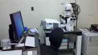
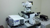
Facility Images
Background Information
Confocal laser scanning microscopy (CLSM or LSCM) is a technique for obtaining high-resolution optical images.[1] The key feature of confocal microscopy is its ability to produce in-focus images of thick specimens, a process known as optical sectioning. Images are acquired point-by-point and reconstructed with a computer, allowing three-dimensional reconstructions of topologically-complex objects. The principle of confocal microscopy was originally patented by Marvin Minsky in 1957,[2] but it took another thirty years and the development of lasers for CLSM to become a standard technique toward the end of the 1980s. (source: Wikipedia)
Documents
LSM 710 Manual (PDF)
Introduction to Confocal Microscopy (PDF)
Confocal Microscopy Guide (PDF)
Equipment Specifications
Please note: More detailed information about the system can be obtained from our staff.
Services Offered:
Confocal Fluorescent Imaging in 4 channels including Zeiss Meta spectral detector.
Brightfield and DIC Imaging (using a transmitted light detector)
User Training.
Data analysis and post-processing.
Excitation sources:
Diode Laser (405 nm) - 30mW
Ar Laser (458/488/514 nm) - 25mW
DPSS 561 - 15mW
Hene Laser (594nm) - 2mW
Hene Laser (633nm) - 5mW
X-Cite 120 Series Illumination system
Detectors:
Zeiss Meta (QUASAR) detector with step size 3.2 - 38.9 nm.
Stand:
Observer.Z1 inverted with motorized focusing drive, fully motorized stage, definite focus system.
FL Filters (visual observation):
Combination of filter sets allowing for simultaneous use with dyes excited in:
Blue spectral range - 458/488nm (GFP,CFP,LUCIFER YELLOW, BCECF, FITC, CY2, ACRIDINE ORANGE)
Green Spectral Range - 543nm (DSRED,RHODAMINE (TRITC), CY3, TEXAS RED, PROPIDIUM IODIDE
Red Spectral Range - 633nm (CY5,DID,ALEXA FLOUR 633)
Violet Spectral Rnage - 405nm (DAPI,HOECHST,AMCA,BFP)
Optics:
Eyepiece PL 10x/23 focusing
EC Plan-Neofluar 10x/0.3 NA
Plan Apo 20x/0.8NA DRY DIC
C-Apochromat 40x/1.2 Water Korr
EC plan neofluar 40x/1.3NA OIL DIC
Plan apo 63x/1.4NA OIL DIC
DIC prism II
DIC prism III
analyzer D/DIC
polarizer D ROT removable
Software:
Zeiss Zen 2011 64 bit Black Edition
Additional Resources:
Dyes: https://www.chroma.com/spectra-viewer
Fluorescence: http://www.thermofisher.com/us/en/home/support/tutorials.html#vid1
Last Updated: 04/01/2024 10:47